March 2009 Archives

Professor K. Tsuji (Osaka City University, Japan) and his colleagues recently reported an interesting application of grazing-exit micro X-ray fluorescence to the analysis of a leaf of Camellia hiemalis. In their experiments, a polycapillary X-ray lens was used to observe a 30 μm area. Grazing-exit geometry was employed to enable the observation of the near surface of the plant. For more information, see the paper, "Grazing Exit Micro X-ray Fluorescence Analysis of a Hazardous Metal Attached to a Plant Leaf Surface Using an X-ray Absorber Method", T. Awane et al., Anal. Chem., 81, 3356-3364 (2009).

Our lab signed a Memorandum of Understanding for research collaboration on X-ray spectrometry and advanced metrology using synchrotron radiation with the Physikalisch-Technische Bundesanstalt (PTB), Germany. The two institutions agreed to promote exchanges of researchers, information, publication of results of the research, and implementation of cooperative research. PTB has several dedicated beamlines at BESSY, Germany's 3rd generation synchrotron radiation facility, and also owns a compact storage ring for photon metrology. Both sides understand the significance of advanced metrology with X-rays for advanced future sciences.

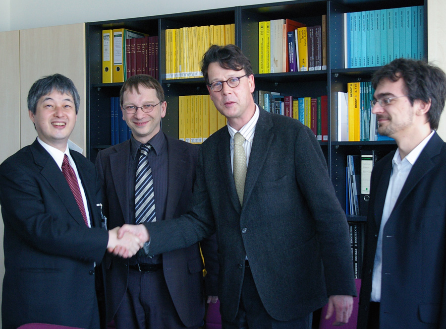
From right to left: Mr. Matthias Muller (PTB, Ph.D. student, X-ray Spectrometry group), Prof. Mathias Richter (PTB, Head of Department, X-ray Metrology using Synchrotron Radiation), Dr. Burkhard Beckhoff (PTB, Head of X-Ray Spectrometry group), Dr. Kenji Sakurai (NIMS, Group Leader, Quantum Beam Center).

At the Photon Factory, KEK, Japan, Dr. T. Okuda (University of Tokyo) and his colleagues have developed a new technique for determining the identity of groups of individual atoms. Scanning tunneling microscopy (STM) is an existing powerful characterization method, which can detect the atomic positions in real space. In order to upgrade the STM by giving it the capability to distinguish chemical species, the research group employed synchrotron X-rays, which excite core-level electrons in the sample's atoms. In this way, secondary electrons can be detected by the STM as they tunnel across the gap. The important point here is that the tunneling current depends on the chemical species. Accordingly, the technique provides chemical imaging. The current spatial resolution is around 10 nm. In the present research, Fe and Ni L absorption edges were chosen to control the core-level excitation. The beamline used was BL-13C. For more information, see the paper, "Nanoscale Chemical Imaging by Scanning Tunneling Microscopy Assisted by Synchrotron Radiation", T. Okuda et al., Phys. Rev. Lett. 102, 105503 (2009)

Obituary - Tomoya Arai

Tomoya Arai, a renowned specialist in X-ray fluorescence spectroscopy and an adviser to Rigaku Corporation, has died at the age of 77 in Osaka, Japan. Dr. Arai was born in 1931 in Tokyo. He was a student at Tokyo Metropolitan University. Immediately after finishing school, he joined Rigaku, where he devoted his life to the development of innovative X-ray fluorescence technologies. In 1962, Dr. Arai developed the first automatic sequential wavelength-dispersive spectrometer in Japan. In 1967, he oversaw the commercialization of an on-line X-ray coating thickness gauge. This was an important industrial application of X-ray fluorescence spectroscopy. In 1969, Dr. Arai proposed the use of an end window Rh tube in an X-ray fluorescence spectrometer. This innovation opened up new opportunities in light element analysis. In 1981, he established a way of analyzing boron. In 1998, Dr. Arai obtained a PhD from the University of Tokyo, where his supervisor was Professor Y. Nihei. Dr. Arai was a recipient of the Birks Award at the 2004 Denver Conference. Some of his valuable experiences have been published in scientific journals. One interesting paper is the "Intensity and distribution of background X-rays in wavelength-dispersive spectrometry", X-Ray Spectrometry, 20, 9-22 (1991). Dr. Arai often spoke of the need to take heed of the raw data. Even in his later years, he never stopped looking at the raw data with his own eyes.

Dr. A. von Bohlen (Institute for Analytical Sciences, Germany) and his colleagues recently published an interesting paper on the analysis of nanoparticles prepared on the substrate by grazing incidence X-ray Standing Waves (XSW) and Total Reflection X-ray Fluorescence (TXRF). The influence of coherence length of X-rays from different X-ray sources, the particle form, particle size and distribution are discussed. For more information, see the paper, "The influence of X-ray coherence length on TXRF and XSW and the characterization of nanoparticles observed under grazing incidence of X-rays", A. von Bohlen et al., J. Anal. At. Spectrom., 2009 (advance article) DOI: 10.1039/b811178b

A research team from the National Natural History Museum in Paris and the American Museum of Natural History in New York recently analyzed the 3D structure of a 300-million-year-old brain of a relative of sharks and ratfish at the European Synchrotron Radiation Facility (ESRF). This is the first time that the soft tissue of such an old fossil brain has ever been found, and the sample studied was found in Kansas, United Sates. The study was performed by combined use of absorption microtomography and a new holotomographic approach, which uses phase-contrasts. The analysis indicated that the area where the brain-like structure reaches the surface of the sample reveals a high concentration of calcium phosphate, whereas the surrounding matrix is almost pure calcium carbonate. The mineralization of the brain might be due to the presence of bacteria that covered the brain shortly before decay and induced its phosphatization. For information about the holotomographic approach, see the paper, "Mixed transfer function and transport of intensity approach for phase retrieval in the Fresnel region", P. Guigay et al., Opt Lett., 32, 1617 (2007). For more information on this research, see the paper, "Skull and brain of a 300-million-year-old chimaeroid fish revealed by synchrotron holotomography", A. Pradela et al., Proceedings of National Academy of Science (published online before print March 9, 2009, doi: 10.1073/pnas.0807047106)

The Chemical Heritage Foundation (CHF) has announced that Dr. Alfred Bader (Cofounder of Aldrich Chemical Company, former chairman of Sigma-Aldrich Corporation) has received the 2009 annual Pittcon Heritage Award. Jointly sponsored by the Pittsburgh Conference on Analytical Chemistry and Applied Spectroscopy (Pittcon) and CHF, this award recognizes outstanding individuals whose entrepreneurial careers have shaped the instrumentation community, inspired achievement, promoted public understanding of the modern instrumentation sciences, and highlighted the role of analytical chemistry in world economies. Dr. Bader founded the Aldrich Chemical Company, a fine chemicals company that later would become the Sigma-Aldrich Corporation, the 80th largest chemical company in the United States. In 1995, he published his autobiography, "Adventures of a Chemist Collector", which details his experiences from Nazi-era refugee to chemist magnate to fine arts connoisseur. In 2008, the second autobiography has been published, "Chemistry & Art: Further Adventures of a Chemist Collector".

At the Shanghai Synchrotron Radiation Facility (SSRF) in China, the Hard X-ray Microfocus Beamline (BL15U1) was commissioned satisfactorily from February to March 2009. The beamline is equipped with an in-vacuum undulator. For further information, visit the Web page, http://ssrf.sinap.ac.cn/english/

Monthly Archives

​​

About Us

Conference Info

Powered by Movable Type 7.902.0