November 2009 Archives

Dr. G. J. Havrilla (Los Alamos National Lab., USA; one of the associate editors of X-Ray Spectrometry journal) and his colleague recently published a very interesting report on the analysis of picoliter droplets, which can be now accurately prepared using Hewlett-Packard's extremely sophisticated technology. The research targets application to analytical science, although the instrument is basically designed for inkjet printing and other similar purposes. It has been shown that dried deposits of single and multielemental solutions generated in picoliter volumes are able to be used as references for micro X-ray fluorescence. Evaporation can have a strong influence on extremely small amounts at the picoliter level, but the research group successfully devised the optimal instrumental conditions by monitoring X-ray fluorescence intensity. For more information, see the paper, "Picoliter droplet deposition using a prototype picoliter pipette: Control parameters and application in micro X-ray fluorescence", U. E. A. Fittschen et al., Anal. Chem., 82, 297 (2010).

Ultimate hard X-ray focusing

For many years, substantial effort has been devoted to developing a good mirror for preparing a small X-ray beam. Professor K. Yamauchi (Osaka University, Japan) and his colleagues have recently reported the breaking of the 10 nm barrier for hard X-rays. They employed a combination of two mirrors; the surface of the first mirror is deformable, in order to compensate for figure error of the second mirror. By such an adaptive optical system, the research group attained a beam size of 7 nm at 20 keV. The experiments were done at BL29XUL, SPring-8. For more information, see the papers, "Breaking the 10 nm barrier in hard-X-ray focusing", H. Mimura et al., Nature Physics doi:10.1038/nphys1457; published online: 22 November 2009; corrected online: 2 December 2009.

Dr. I. Han (Ağİbrahim Çeçen University, Turkey) and his colleagues have published a paper on the relationship between the Kβ/Kα X-ray fluorescence intensity ratio and valence-electron configurations. For more information, see the paper, "Valence-electron configuration of Fe, Cr, and Ni in binary and ternary alloys from Kβ-to-Kα X-ray intensity ratios", I. Han et al., Phys. Rev. A80, 052503 (2009).

Foamlike, cellular structures of the monolayer of organic capped nanoparticles can sometimes be observed on liquid surfaces. Professor M. K. Sanyal (Saha Institute of Nuclear Physics, India) and his lab members studied the time evolution in the structure and morphology of transferred monolayers of gold-thiol nanoparticles, formed at the air-water interface at different surface pressure, on to a silicon surface. The research group employed two complementary techniques, X-ray reflectivity and atomic force microscopy (AFM), to see the whole drying-mediated self-assembly of nanoparticles. For more information, see the paper, "Nanopattern formation in self-assembled monolayers of thiol-capped Au nanocrystals", R. Banerjee et al., Phys. Rev. E80, 056204 (2009).

Nanometer scale dipole moments in the polarization clusters in BaTiO3 are believed to be thermally excited and thermally relaxed within a picosecond time scale. However, so far, there have been no reports on the direct observation of the dynamics of these dipole moments in such a very short time scale. The limitation here is mainly due to the low spatial coherence of the X-ray beam, in particular when synchrotron radiation is used as a light source. Professor K. Namikawa (Tokyo Gakugei Univ, Japan) and his colleagues have recently obtained some interesting results. To measure the time correlation of speckle intensities, they employed a soft X-ray pulse laser (7 ps in pulse width, 3.5×10¹⁰ photons/sec/pulse, 13.9 nm in wavelength, band width 10-4, angular spread 0.5 mrad) at Japan Atomic Energy Agency, Kizugawa, Japan, and a Michelson-type delay pulse generator as well as an X-ray streak camera. Spatial coherence in their system was estimated at more than 90 %. The evolution of the relaxation time of the dipole moment near the Curie temperature (TC) was studied. It was found that the maximum relaxation time (~90 ps) appears at a temperature of 4.5 K above the TC, being coincident with the one where the maximum polarization takes place. For more information, see the paper, "Direct observation of the critical relaxation of polarization clusters in BaTiO3 using a pulsed X-ray laser technique", K. Namikawa et al., Phys. Rev. Lett., 103, 197401 (2009).

So far, X-ray microscopy with many types of lens has achieved great success in the observation of biological cells. In order to extend the limits of spatial resolution and efficiency, X-ray diffraction microscopy (also called coherent X-ray diffraction imaging), which uses coherent X-rays and some image reconstruction algorithms instead of an optical lens system, is now considered as a promising procedure to see whole cells at once and pick out much smaller features, down to around 10 nm or even less. A research group led by Professor C. Jacobsen (Stony Brook University, USA) recently reported the results for yeast cells with 520 eV soft X-rays at the Advanced Light Source (ALS) at Lawrence Berkeley National Laboratory, USA. Dr. A. Madsen (European Synchrotron Radiation Facility (ESRF), Grenoble, France) and his colleagues observed the cells of the bacteria D. radioduran with 8 keV X-rays. The advantage of using hard X-rays is the ease of sample handling, and the validity of thin sample approximation for future 3D reconstructions through phasing a diffraction volume. In both cases, a rapid freezing technique (instead of previously used freeze-drying) was used to avoid the effects of radiation damage from synchrotron X-ray photons. The Stony Brook group plunged cells in their natural wet state into liquid ethane and maintained them at below -170 oC, leading to the reduction of artifacts due to damage from dehydration, ice crystallization, and radiation. In the ESRF setup, as absorption in air of 8 keV X rays is small, a nonvacuum environment was implemented for ease of sample handling. Similar to the system for macromolecular crystallography applications, they based the samples in a continuous cryogenic nitrogen gas jet at around -165 oC. The spatial resolution was 25 nm and 30-50 nm, for soft and hard X-rays cases, respectively. For more information, see the papers, "Soft X-ray diffraction microscopy of a frozen hydrated yeast cell", X. Huang et al., Phys. Rev. Lett., 103, 198101 (2009), and "Cryogenic X-ray diffraction microscopy for biological samples", E. Lima et al., Phys. Rev. Lett., 103, 198102 (2009)

Professors T. Narayanan (ESRF, Grenoble, France), M. Giglio (XFEL, Hamburg, Germany) and their collaborators have recently published an interesting paper on a novel method to map the two-dimensional transverse coherence of an X-ray beam. The technique uses the dynamical near-field speckles formed by scattering from colloidal particles, which are executing Brownian motions. It is possible to measure the change of the interference fringes, and consequently the fluctuation of speckles. It was found that the coherence properties of synchrotron radiation from an undulator source are obtained with high accuracy. For more information, see the paper, "Probing the transverse coherence of an undulator X-ray beam using Brownian particles", M. D. Alaimo et al., Phys. Rev. Lett., 103, 194805 (2009).

The recipient of the 4th Asada Award, which is presented in memory of the late Professor Ei-ichi Asada (1924-2005) to promising young scientists in X-ray analysis fields in Japan, is Dr. Akiko Hokura (Tokyo Denki Univ., "Study on accumulation of heavy metals in phytoremediation plant by synchrotron radiation micro XRF imaging and XAFS analysis"). From this year, the Discussion Group of X-ray Analysis, the Japan Society for Analytical Chemistry decided to establish the special award to recognize scientists who exhibit outstanding achievement and make a substantial contribution to the advancement of the X-ray analysis field. The recipient of the special award 2009 is Dr. Toshio Shiraiwa, who contributed greatly in the early days of X-ray absorption spectroscopy by means of his short-range order theory ("The theory of the fine structure of the X-ray absorption spectrum", J. Phys. Soc. Jpn. 13, 847 (1958)) and also provided the basis of the fundamental parameter method in X-ray fluorescence by Fujino-Shiraiwa's formula ("Theoretical calculation of fluorescent X-ray intensities in fluorescent X-ray spectrochemical analysis", Jpn. J. Appl. Phys. 5, 886 (1966)) The ceremony was held during the 45th Annual Conference on X-Ray Chemical Analysis, Japan, at Osaka City University, Osaka.

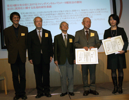
From right to left: A. Hokura, T. Shiraiwa, S. Ikeda, H. Wakita and H. Hayashi.

The discovery of X-rays was named the most important modern scientific achievement in a poll conducted for the Science Museum London, beating the Apollo spacecraft and DNA. Nearly 50,000 members of the public voted in the museum or online. The emblem of the London museum's centenary is now an X-ray machine. For further information, visit the museum's Web page, http://www.sciencemuseum.org.uk/

Monthly Archives

​​

About Us

Conference Info

Powered by Movable Type 7.902.0