Kenji Sakurai
|
Kenji Sakurai Director, Group Leader, X-Ray Physics Group, National Institute for Materials Science
Adjunct Professor at Charles University in Prague, Czech
|
|
||||||||||||||||||||||||||||||||||||||||||||||||||||||||||||||||||||
Why X-rays? As you well know, X-rays give atomic-scale structure information as an average for quite a large area - you can always see the whole forest even if you are only looking at the trees, which is not the case for other major microscopic tools. Another advantage is that X-rays can be used for analyzing thick, voluminous materials as well as the surface and interfaces of thin films. In addition, sample preparation is easy and, in most cases, you can use your sample as is. Measurement can be done in air, and even in liquid. This freedom in the measurement environment is very attractive. Furthermore, X-ray analysis is generally quantitative and reproducible. Because of this, X-ray techniques are reliable and widely used in industrial and medical applications. However, the biggest advantage of X-ray analysis is its non-destructive nature. This ensures that the same specimen can be measured using other techniques after X-ray measurement. So, what do you think? X-rays are a really attractive tool for analysis, aren't they? However, our research is concerned with what has not yet been achieved by X-rays, rather than attractive applications of the feasible using established tools. We are interested in developing novel analytical methods (theoretical as well) and instruments which will be useful for future materials research. The methods are always immature in the early stages of the research. But that is the nature of science. We prefer to choose such new topics, which are not so easy to understand in terms of study techniques. Instrumentation is key to opening up a new field. In addition to the novelty of the experimental methods, we always pay due respect to careful, in-depth and high-quality data analysis based on original mathematical procedures.
(1) X-Ray Spectrometry and Imaging for Future Materials Research
X-ray fluorescence spectroscopy can be used not only for the identification/quantification of elements but also for chemical speciation. Our lab is interested in the potential feasibility of Kβ spectra for 3d transition elements, measured with a middle energy resolution around 10 eV, with the main idea being the efficient detection of intensity changes of Kβ′ and Kβ5 rather than precise measurements of energy shifts that require high energy-resolution. Though our earlier experiments were performed at the SPring-8, we are now developing a competitive spectrometer for laboratory use. Lanthanides' Lβ and L g spectra are other interesting targets. Micro X-ray fluorescence imaging is a promising method for obtaining positional distribution on specific elements in a non-destructive manner. So far, the technique has usually been performed by a 2D positional scan of a sample against a collimated beam. However, the total measuring time can become quite long, since a number of scanning points are needed in order to obtain a high-quality image. Our lab has developed a completely different way of performing imaging of elements much more quickly. A combination of grazing-incidence geometry using a rather wide beam and parallel optics for detecting X-rays can produce an X-ray fluorescence image with 1 M pixels and with 15 μm resolution in 30-100 msec. The technique has the potential to open up new frontiers in X-ray imaging, particularly in element-selective movie applications. The technique has been extended to X-ray absorption fine structure (XAFS), X-ray diffraction and Compton X-ray scattering etc.
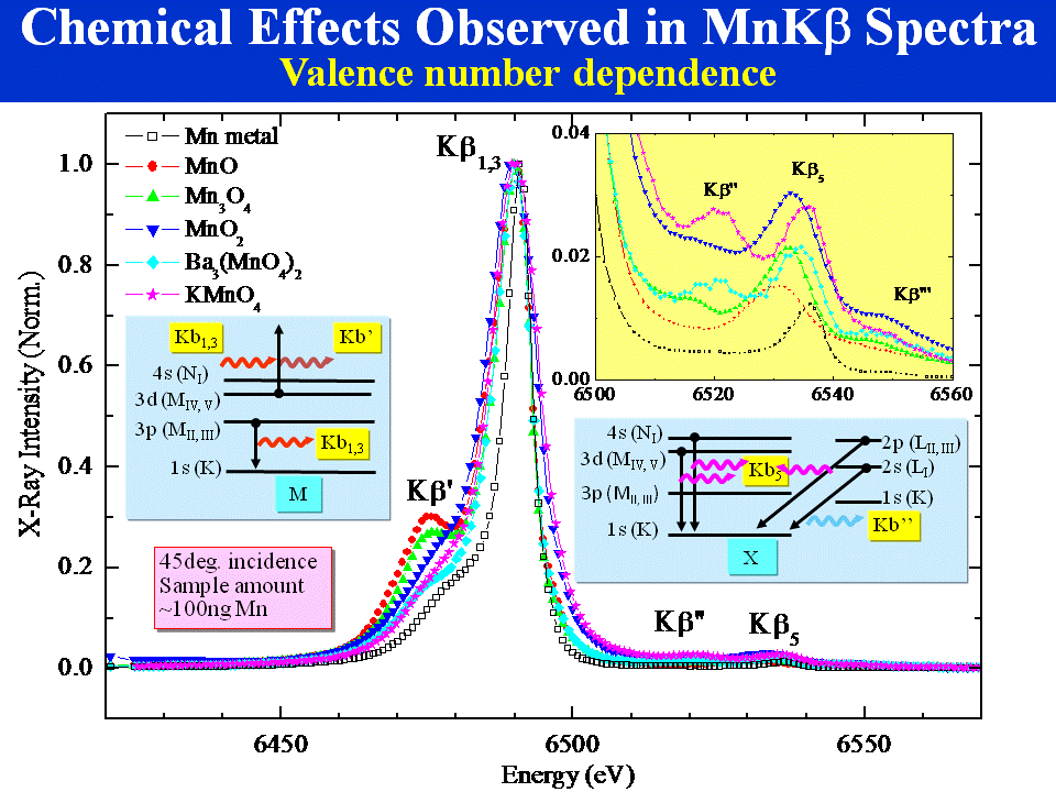 Chemical Characterization
 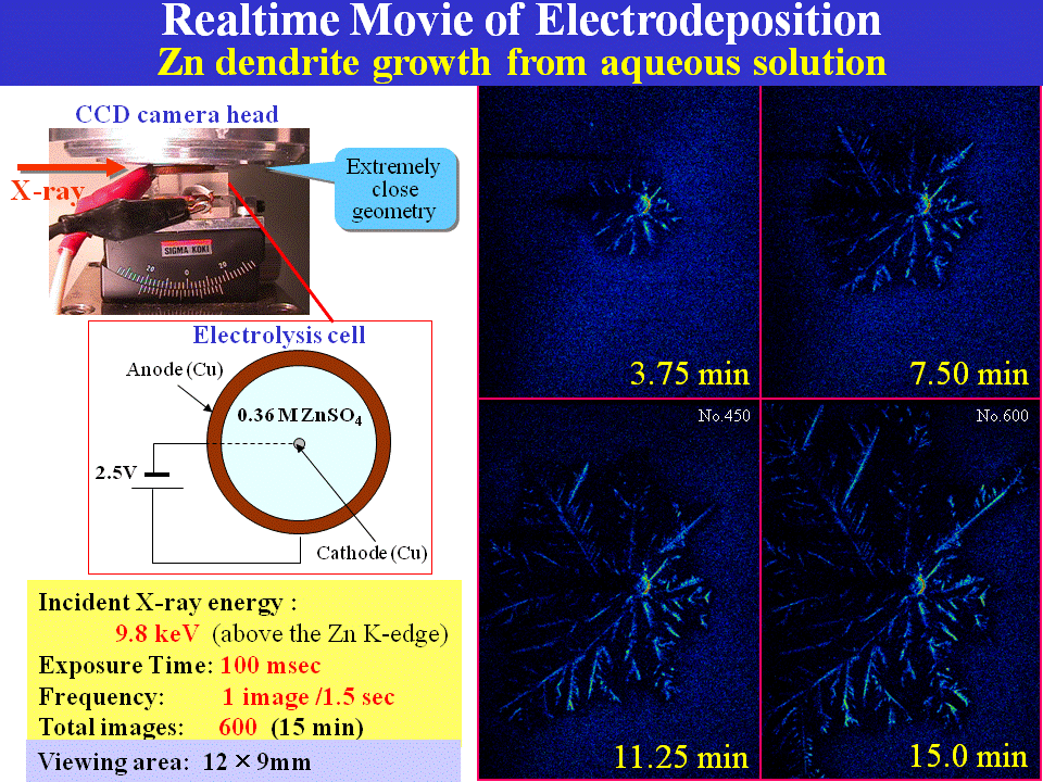 Fast Imaging
(2) Buried Interface Sciences by X-Rays and Neutrons
There are increasing demands for sophisticated metrology in order to observe multilayered materials with nano-structures (dots, wires, etc), which are finding applications in electronic, magnetic, optical and other devices. Unlike many other surface-sensitive methods, X-ray and neutron analysis is known for its ability to see even 'buried' function interfaces as well as the surface. It is highly reliable in practice, because the information, which ranges from the atomic to mesoscopic scale, is quantitative and reproducible. However, we now realize that the method should be upgraded further to cope with more realistic problems in nano sciences and technologies. Current X-ray methods can give atomic-scale information for quite a large area on a scale of mm2-cm2. These methods can deliver good statistics for an average, but sometimes we need to be able to see a specific part in nano-scale rather than an average structure. In addition, there is a need to see unstable changing structures and related phenomena in order to understand more about the mechanism of the functioning of nano materials. Quick measurements are therefore important. Furthermore, in order to apply the method to a more realistic and complex system, we need some visual understanding to discuss the relationship among the different structures that are present in the same viewing. Therefore, 2D/3D real-space imaging is important. Interpretation of roughness is another significant subject, while combination with grazing-incidence small angle scattering (GISAS) will become much more widespread than before. The use of coherent beams and several other new approaches are significant in this respect. Developing an analytical procedure that does not depend on the model is also extremely important.
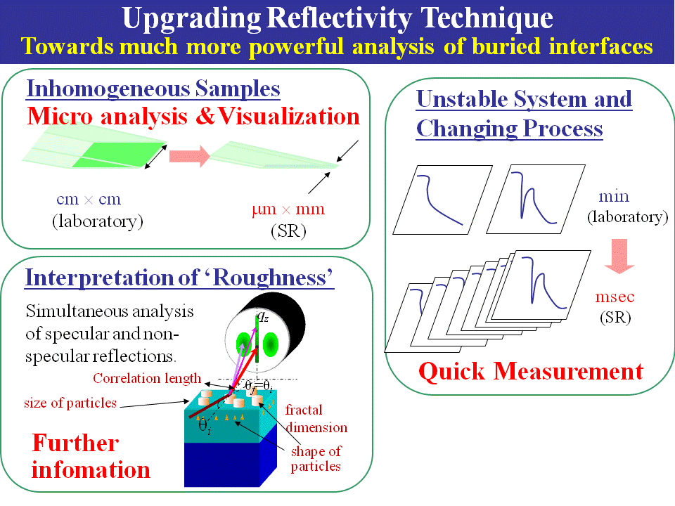 Instrumentation for Advanced Reflectometry
(3) Contribution to Energy, Environment and Safety Problems through X-Ray Technologies
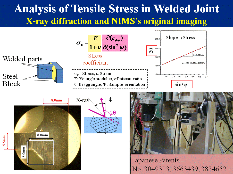
We are eager to make use of our X-ray techniques for unresolved social problems. So far, we have been involved in the non-destructive evaluation of hydrogen tanks for fuel cell cars and welded joints in nuclear plants by X-ray transmission and X-ray diffraction stress analysis, respectively.



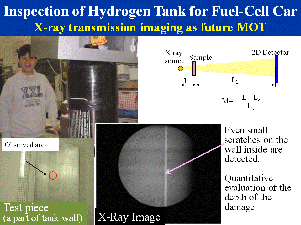 Non-destructive inspection
 Stress analysis